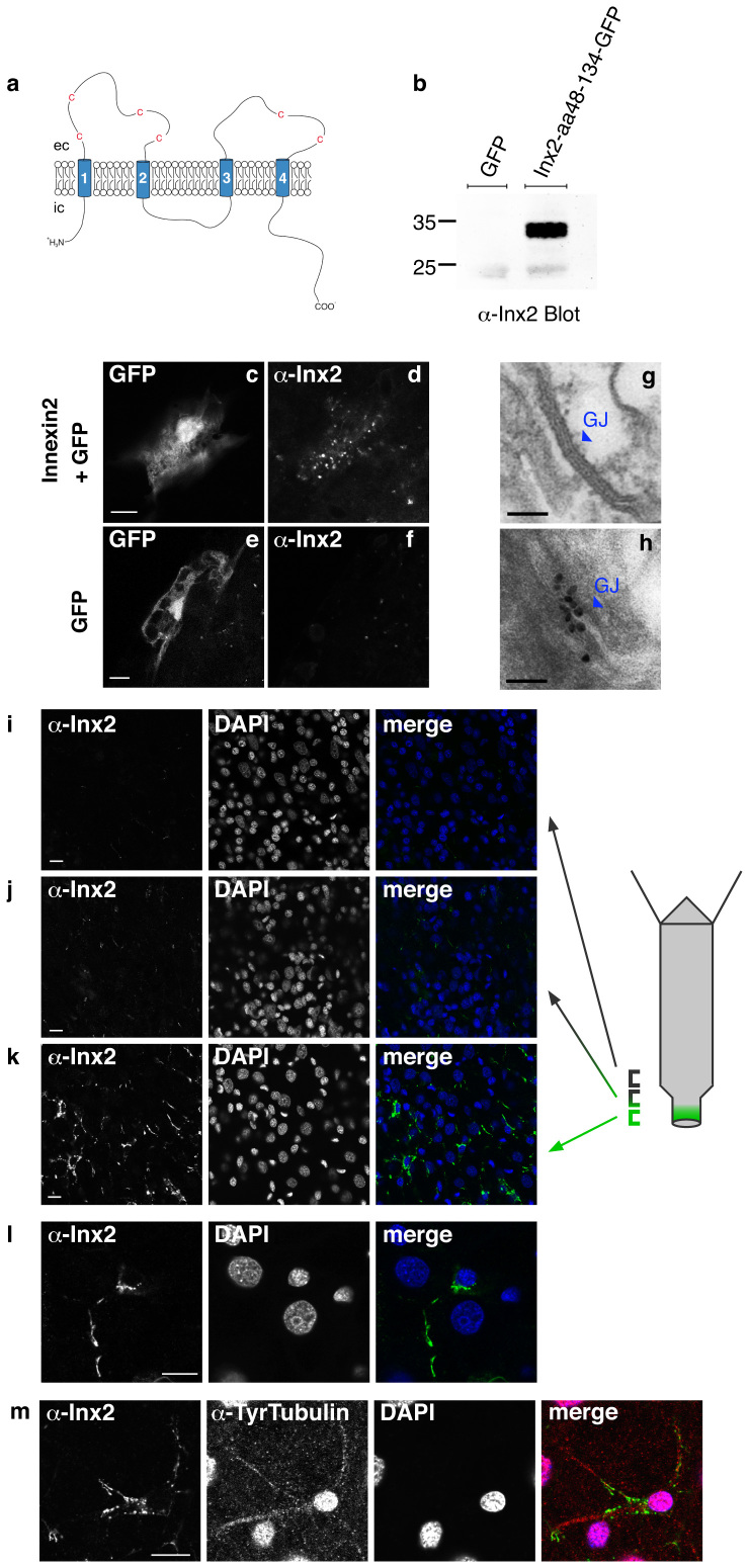
Figure 2. Immunofluorescent staining of innexin-2 in gap junctions in Hydra.
(a) Schematic drawing of the predicted structure of hydra innexin showing four transmembrane domains and conserved cysteine residues in the extracellular (ec) loops. N- and C-terminus are intracellular (ic). The first extracellular loop of innexin-2 (aa48–134) was used for antibody production. (b) Purified recombinant GFP-tagged innexin-2 (aa48–134) from E.coli was detected by the innexin-2 antibody at the appropriate size (ca. 35 kD) in immunoblot. The antibody did not detect GFP. (c–f) Ectopic expression of GFP (used as a transfection marker) and untagged innexin-2 in hydra epithelial cells transfected with the particle gun. To visualize innexin-2 expression, animals were fixed and stained with innexin-2 antibody 48 hours post-transfection. Transfected GFP-expressing epithelial cells displayed a punctate innexin-2 pattern in immunofluorescence when co-transfected with innexin-2 (d). Control animals transfected with GFP only have no detectable innexin-2 signal (f). Scale bar: 10 μm. (g) and (h) Immunogold staining of innexin-2 gap junction in peduncle tissue. (g) TEM image of a typical gap junction (enlarged from Fig. 1A); (h) immunogold staining of innexin-2 gap junction in peduncle tissue. Scale bar: 100 nm. (i–k) Immunostaining of whole animals with innexin-2 antibody revealed innexin-2 positive green spots primarily in the peduncle region (k); more apical areas in the peduncle showed decreased amounts of antibody staining (i and j); the gastric region contained no innexin-2 positive green spots. (l) High magnification image of an innexin-2 positive cell in the peduncle region. The innexin-2 positive green spots were often clustered as strings along nerve processes. Nuclei stained with DAPI. Projections of confocal images covering a depth of 2–3 μm. Scale bar: 10 μm. (m) Co-immunostaining of hydra with innexin-2 antibody and tyrosine-tubulin antibody (Sigma) showed that innexin-2 staining in nerve cells was localized along nerve cell processes. The anti-tyrosine-tubulin staining in nerve cell nuclei is regularly observed but appears to be an artifact (see also Figure 3). Nuclei stained with DAPI. Projections of confocal sections covering a depth of 2 μm. Scale bar: 10 μm.

