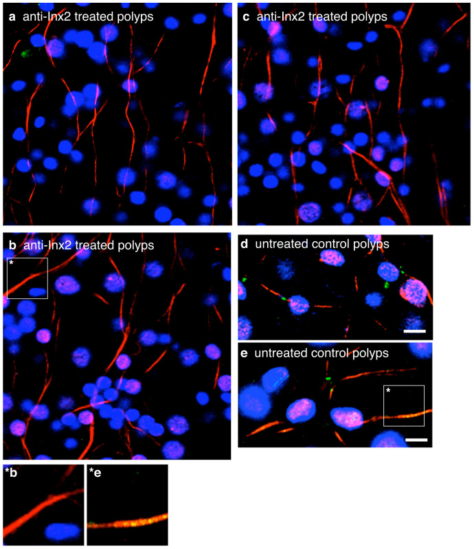
Figure 3. Treatment of Hydra polyps with innexin-2 antibody eliminated innexin-2 stained gap junctions in peduncle tissue.
(a), (b) and (c) Confocal images of three anti-innexin-2 treated polyps fixed after 3 days and co-immunostained with innexin-2 antibody (green) and tyrosine-tubulin antibody (red). Nuclei are stained with DAPI (blue). (d) and (e) Confocal images of two DMSO treated control polyps. Note that innexin-2 spots (green) are yellow where directly overlapping with strong anti-tubulin (red) stained processes. (*b and *e) Enlargements of single nerve cell processes from b (anti-innexin-2 treated polyps) and e (untreated control polyps). Projections of confocal images covering a depth of 2 μm. Scale bar, 10 μm.

