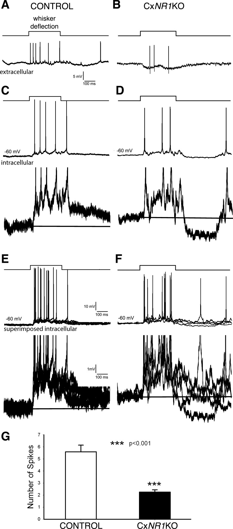
Fig. 6.
Comparison of tonic responses to whisker deflection between anesthetized control and CxNR1KO mice. A and B: extracellular recordings. C and D: sharp electrode intracellular recordings show different spike numbers (top traces) and difference in membrane depolarization (10 times enlarged bottom traces) between control and CxNR1KO mice. E and F: superimposed 5 traces show that in control mice the depolarization is sustained, whereas in CxNR1KO mice the depolarization is not stable (bottom traces). G: averaged spike numbers during whisker deflection are significantly deferent between control and CxNR1KO mice.

