
Fig. 1.
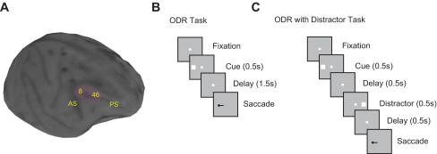
A: structural MRI of 1 adolescent monkey brain. The shaded areas indicate the recording sites in the dorsolateral prefrontal cortex (PFC). Black spots in the image are artifactual “shadows” created by ceramic screws in the skull. AS, arcuate sulcus; PS, principal sulcus. B: successive frames indicate the sequence of events in the oculomotor delayed response (ODR) task. Monkeys were required to remember the stimulus location and to saccade to it after a delay period. B: sequence of events in the ODR with Distractor task. The presentation of the stimulus is the same as in the ODR task, but 1 distractor is shown in the middle of the delay period.