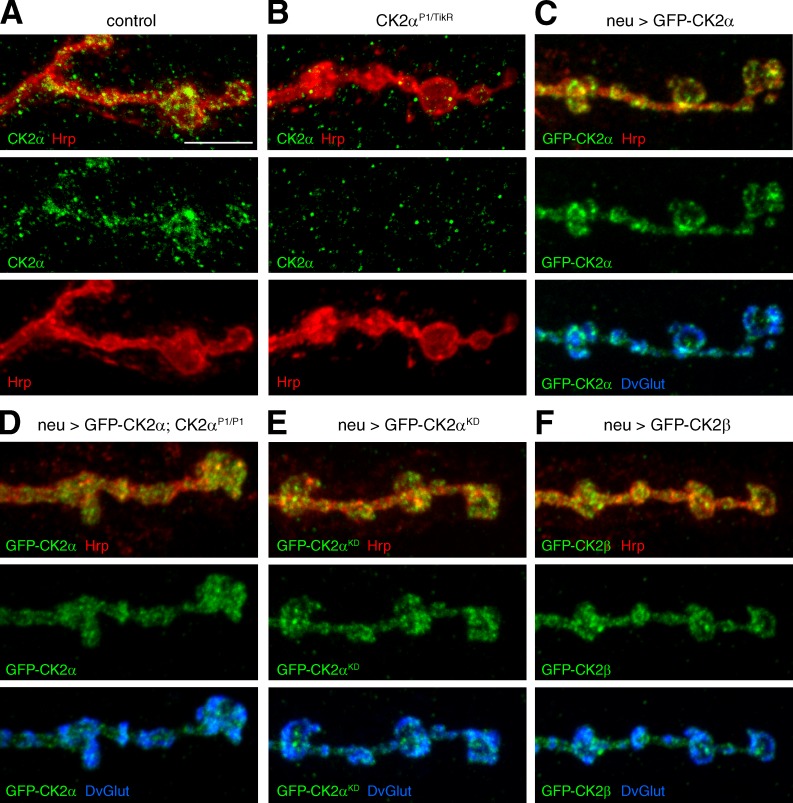
Figure 5.
CK2α and CK2β localize to the presynaptic nerve terminal. (A and B) Analysis of endogenous CK2α localization. CK2α (green) is present within the presynaptic nerve terminal marked by Hrp (red; A). In CK2αP1/TikR mutant animals presynaptic CK2α staining is absent (B). (C–F) Analysis of the synaptic localization of GFP-tagged CK2α and CK2β. (C) GFP-tagged wild-type CK2α (green) localized to synaptic bouton and interbouton regions marked by the synaptic vesicle marker DvGlut (blue). (D) A comparable distribution was observed when GFP-CK2α was expressed in CK2α mutant animals. GFP-tagged CK2α completely restored synapse stability in these animals, indicating functional distribution. (E) Presynaptic CK2α localization did not depend on kinase activity. (F) Neuronal expression of GFP-tagged CK2β (green) resulted in a similar presynaptic localization. Bar, 5 µm.

