Figure 1.
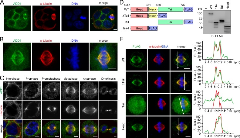
Association of ADD1 with mitotic spindles via its head domain. (A) MDCK cells were allowed to form cell colonies and were then stained for ADD1, α-tubulin, and DNA. Note that one of the cells in the colony is undergoing mitosis. (B) HeLa cells were stained for ADD1, α-tubulin, and DNA. Representative images from confocal z-stack projections are shown. (C) HeLa cells were stained for ADD1, α-tubulin, and DNA. Representative confocal images show the association of ADD1 with mitotic spindles throughout mitosis and with midzone microtubules during cytokinesis. (D) FLAG epitope–tagged ADD1 (FLAG-ADD1) and mutants were transiently expressed in HEK293 cells and were analyzed by immunoblotting (IB) with anti-FLAG. (E) HeLa cells transiently expressing FLAG-ADD1 and mutants were stained for FLAG-ADD1, α-tubulin, and DNA. (left) Representative confocal images from the cells in metaphase are shown, which were from a single experiment out of three repeats (n > 140). (right) Graphs show the relative fluorescence intensity (F.I.) at the lines that were scanned by confocal microscopy. a.u., arbitrary unit. Bars, 5 µm.

