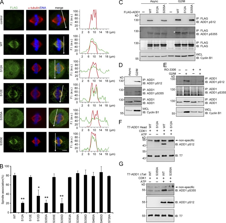
Figure 2.
Phosphorylation of ADD1 at Ser12 and Ser355 by CDK1 is crucial for ADD1 association with mitotic spindles. (A) HeLa cells transiently expressing FLAG-ADD1 and mutants were stained for FLAG-ADD1, α-tubulin, and DNA. (left) Representative confocal images from the cells in metaphase are shown, which were from a single experiment out of three repeats (n > 160). (right) Graphs show the relative fluorescence intensity (F.I.) of the lines that were scanned by confocal microscopy. a.u., arbitrary unit. Bar, 5 µm. (B) The percentage of FLAG-ADD1 association with mitotic spindles in the total number of mitotic cells counted was measured (n > 160). Values (means ± SD) are from three independent experiments. *, P < 0.05; **, P < 0.01. (C) FLAG-ADD1 and mutants were transiently expressed in HEK293 cells. The cells were synchronized in the G2/M phase or remained asynchronized (Async) before they were lysed. FLAG-ADD1 and mutants were immunoprecipitated (IP) by anti-FLAG, and the immunocomplexes were analyzed by immunoblotting (IB) with antibodies to ADD1 pS12 or pS355. S355-phosphorylated ADD1 is indicated by a white asterisk. WCL, whole-cell lysates. (D) HeLa cells were synchronized in the G2/M phase or remained asynchronized before they were lysed. Endogenous ADD1 was immunoprecipitated by anti-ADD1, and the immunocomplexes were analyzed by immunoblotting with antibodies to ADD1 pS12 or pS355. (E) HeLa cells were synchronized in the G2/M phase (+) or remained asynchronized (−). The cells were treated with 10 µM of the CDK1 inhibitor RO-3306 for 1 h before they were lysed. Endogenous ADD1 was immunoprecipitated by anti-ADD1, and the immunocomplexes were analyzed by immunoblotting with antibodies to ADD1 pS12 or pS355. S355-phosphorylated ADD1 is indicated by a white asterisk. (F and G) Purified T7-tagged ADD1 head domain (T7-ADD1 head) or the mutant with a deletion of the tail domain (Δtail) were incubated with recombinant CDK1 in the presence (+) or absence (−) of ATP for 30 min. The phosphorylation of ADD1 at S12 and S355 was analyzed by immunoblotting with antibodies to ADD1 pS12 or pS355.

