Figure 3.
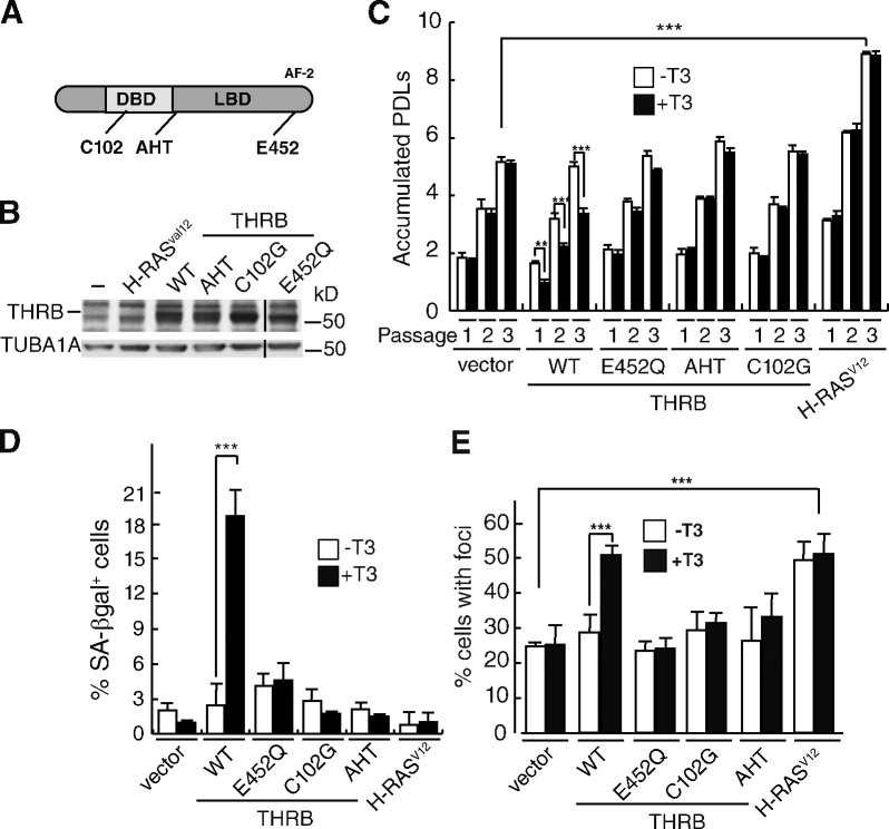
Transcriptionally active THRB mediates TH-induced senescence and DNA damage in MEFs. (A) Schematic representation of THRB, showing positions of the mutations C120G, AHT, and E452Q. DBD, DNA-binding domain; LBD, ligand-binding domain. (B) Thra/ThrbKO MEFs were transduced with vectors for the different mutants or H-RasV12, and after selection, THRB was detected by Western blotting. TUBA1A was used as a loading control. Black lines indicate the removal of an intervening lane for presentation purposes. (C) PDLs, estimated after one, two, and three passages (P < 0.0001, n = 3). (D) Percentages of SA-βgal+ cells after passage 3. (E) Percentage of cells having DNA damage foci determined from immunofluorescence of γ-H2AFX and TP53BP1 and merged images (P < 0.0001, n = 3). Results are presented as means ± SD. **, P < 0.01; ***, P < 0.001. WT, wild type.

