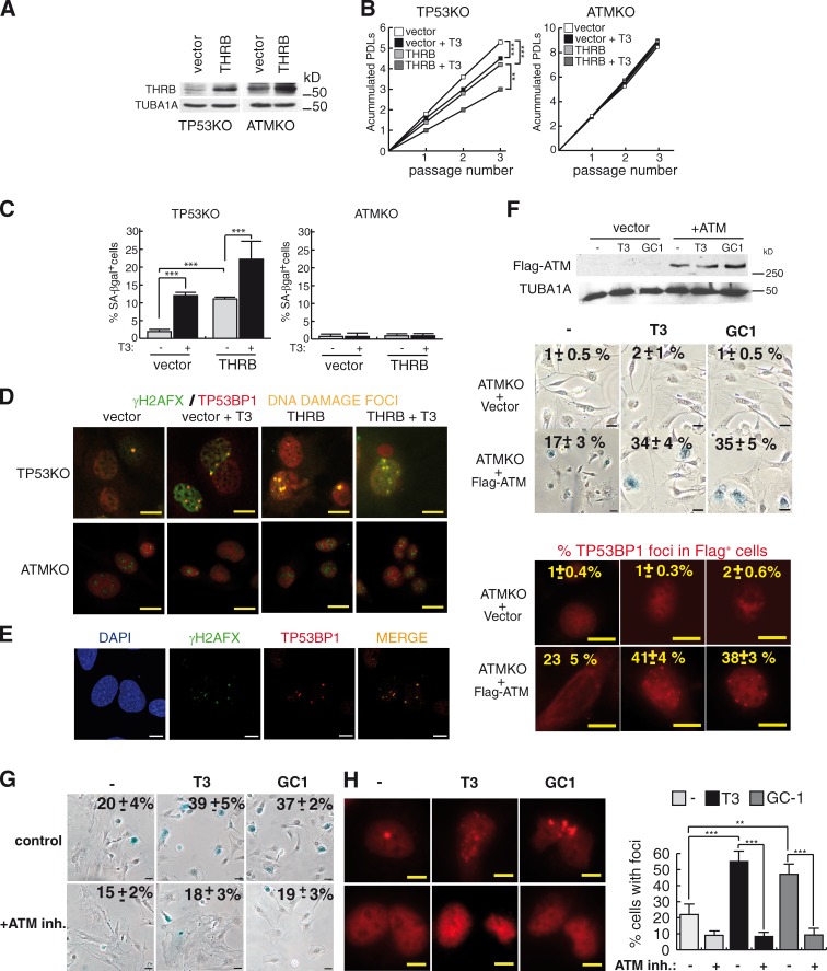
Figure 5.
THRB does not induce senescence in ATMKO MEFs. (A) THRB levels after transduction of an empty vector or the receptor in TP53- and ATM-deficient cells. TUBA1A was used as a loading control. (B) Accumulated PDLs in the presence and absence of T3 at three consecutive passages after selection (P < 0.0001, n = 3). (C) SA-βgal+ cells at passage 3 (P < 0.0001, n = 3). (D) Representative merge images of double immunofluorescences of γ-H2AFX and TP53BP1 in TP53 and ATMKO MEFs incubated with and without T3 during three passages. (E) Confocal microscopy images of γ-H2AFX and TP53BP1 in TP53KO MEFs. (F) ATM levels in ATMKO MEFs transfected with an empty vector or Flag-ATM. Percentages of SA-βgal+ cells after one passage in T3 and GC-1–treated cells, and representative images of TP53BP1 cells, indicating the percentages of Flag-positive cells with foci, are shown at the bottom. (G and H) Percentages of SA-βgal+ cells (G) and TP53BP1 foci (H) in wild-type MEFs pretreated with the ATM inhibitor KU-55993 (10 µM) for 2 h and with T3 or GC-1 for one passage. Bars: (D) 10 µm; (E, F [bottom images], and H) 10 µM; (F [top images] and G) 20 µM. Results are presented as means ± SD. **, P < 0.01; ***, P < 0.001. inh., inhibitor.

