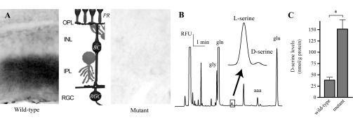
Fig. 1.

A lack of d-amino acid oxidase (DAO) activity results in an increase in d-serine. A: a horseradish peroxidase (HRP)-3–3′ diaminobenzidine (DAB) reaction product, the end product in a DAO activity assay, was visible in the inner plexiform layer (IPL) of wild-type retina but not the ddY/DAO− mutant retina. The middle shows a schematic of the basic retinal circuitry. The main 3-neuron circuit is presented in black: with light response flowing from photoreceptor, PR; to bipolar cell, BC; to retinal ganglion cell, RGC. OPL, outer plexiform layer; INL, inner nuclear layer. B: CE, capillary electrophoresis. An electropherogram, time vs. relative fluorescent units (RFU) showing the separation of l- and d-serine from homogenized mouse retina. Also noted: gly, glycine; gln, glutamine; aaa, aminoadipic acid; glu, glutamate. C: cumulative results of d-serine levels measured from homogenized wild-type and mutant retinas normalized to protein levels. *P < 0.05 between genotypes.
