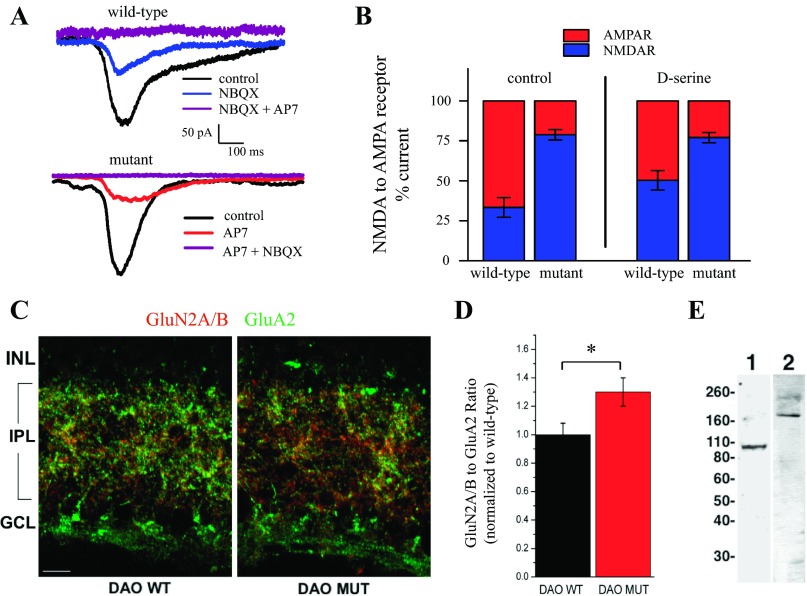
Fig. 5.
NMDA receptor component of the light response is greater in ddY/DAO− mice. A: the combination of AP7 and 2,3-dioxo-6-nitro-1,2,3,4-tetrahydrobenzo[f]quinoxaline-7-sulfonamide disodium salt (NBQX) completely blocks current responses of RGCs in wild-type and mutant mice. B: the percentage of the current mediated by NMDA (blue) and AMPA (red) receptors (R) under different conditions. The left side represents the percentage of currents recorded in control Ringer for both wild-type and mutant mice. The right side compares the percentages in relation to a Ringer solution containing 100 μM d-serine to maximize the NMDA receptor contribution. C: retina cryosections from DAO wild-type (DAO WT) and DAO mutant (DAO MUT) mice were double-labeled by immunofluorescence for GluN2A/B (red) and GluA2 (green) receptor subunits. GCL, ganglion cell layer. The scale bar in C represents 10 μm. D: changes in the ratio of GluN2A/B to GluA2 immunofluorescence of mutant animals compared with their wild-type counterparts (DAO WT, n = 7; DAO MUT, n = 7). *P < 0.05 between genotypes. E: Western blots of mouse retinal proteins probed with antibodies against GluA2 (lane 1) and GluN2A/B (lane 2). The molecular weights (in kilodaltons) and position in the gel of protein standards are indicated to the left.

