Figure 1.
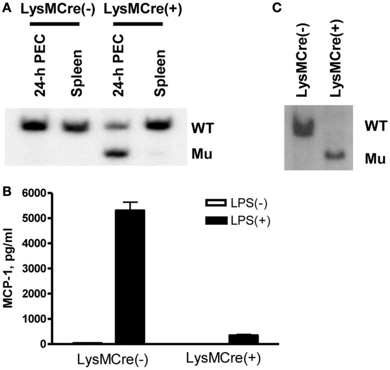
Generation of myeloid cell-specific MCP-1-deficient mice. (A) Genomic DNA was isolated from 4-day TG-induced PEC or spleen of LysMCre−, MCP-1flox/flox or LysMCre+, MCP-1flox/flox mice. The DNA were digested with PstI and subjected to Southern blotting for the presence of WT or mutant (Mu) allele. (B) 1.8 × 10⁶ PEC from LysMCre−, MCP-1flox/flox or LysMCre+, MCP-1flox/flox mice were cultured in 1 ml medium for 24 h in the absence or presence of 100 ng/ml LPS. The concentration of MCP-1 in the culture supernatants was measured by ELISA. Data is presented as the mean ± SD obtained with cells from three mice. (C) PEC isolated 4 days after TG injection were incubated in tissue culture plates at 37C overnight, non-adherent cells were removed, and then adherent cells were lysed to obtain genomic DNA. The DNA were digested with PstI and then subjected to Southern blotting for the presence of WT or mutant (Mu) allele.

