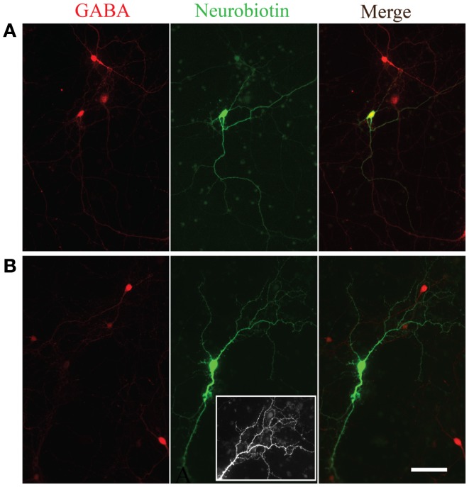
Figure 5.
Morphological evidence for GABAergic and glutamatergic neurons. GABA immunoreactivity (red, left panels) in two neurons filled with Neurobiotin (green, middle panels). The overlay of the two channels is shown in the right panels. Note the smooth appearance of processes from the neuron immunopositive for GABA (A) compared to the spiny appearance of the cell immuno-negative for GABA (B). Inset in the bottom middle panel is an expanded view of the spiny processes on the dendritic tree of the neuron immuno-negative for GABA.

