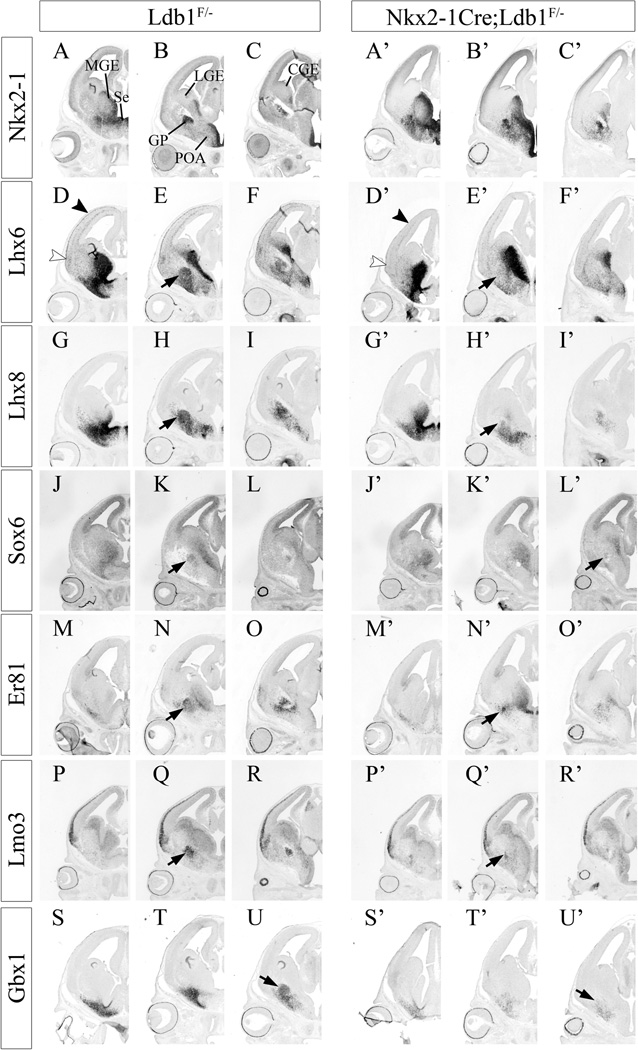
Fig. 6.
Expression of genes that regulate and/or mark MGE development are reduced in conditional Ldb1 mutants, as determined by in situ hybridization at E14.5. Coronal telencephalic hemisections compare gene expression between control (Ldb1+/−; left) and mutant (Nkx2.1-Cre+; Ldb1f/−; right). Three planes of section are shown, with rostral at the left and caudal at the right. Nkx2.1 (A–C, A’–C’); Lhx6 (D–F, D’–F’), black arrowhead shows reduced Lhx6+ neocortical cortical interneurons in mutant; white arrowhead shows reduced Lhx6+ paleocortical cortical interneurons in mutant; black arrow shows reduced GP in mutant; Lhx8 (G–I, G’–I’), black arrow shows reduced GP in mutant; Sox6 (J–L, J’–L’), black arrow shows reduced GP in mutant; Er81 (Etv1; M–O, M’–O’), black arrow shows reduced GP in mutant; Lmo3 (P–R, P’–R’), black arrow shows reduced GP in mutant; Gbx1 (S–U, S’–U’); black arrow shows reduced GP in mutant. Abbreviations: CGE: caudal ganglionic eminence, GP: globus pallidus, LGE: lateral ganglionic eminence, MGE: medial ganglionic eminence, POA: preoptic area, Se: septum.

