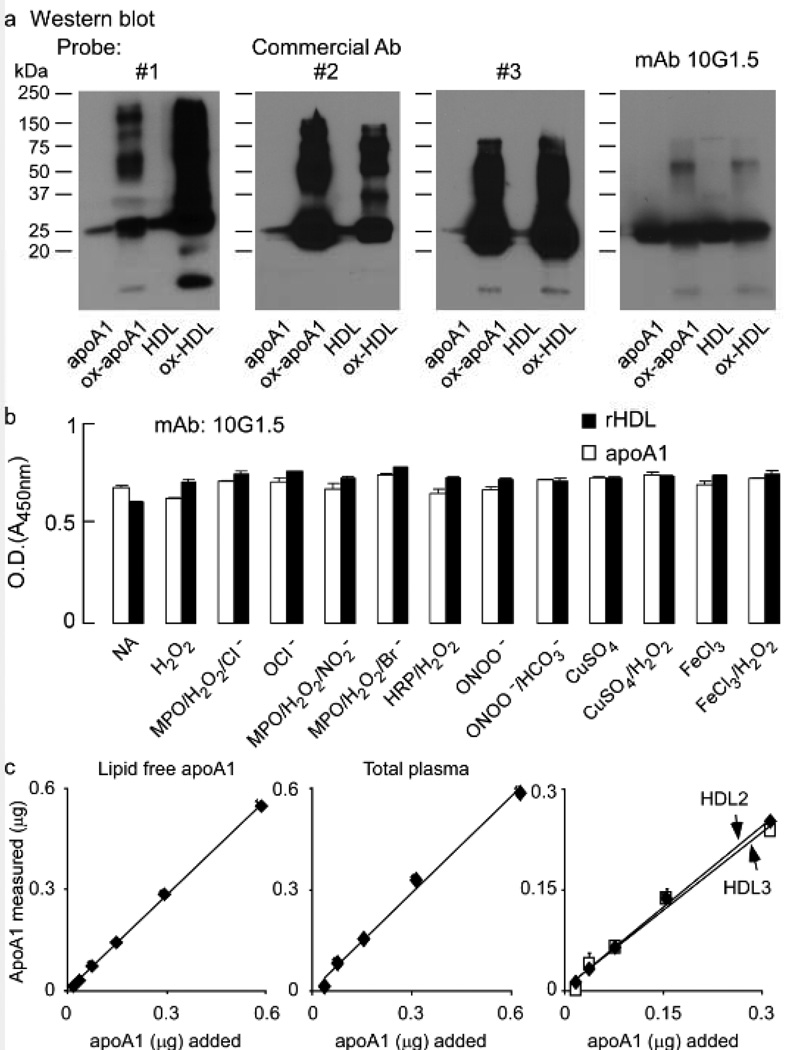
Figure 1.
ApoA1 or apoA1 in reconstituted HDL were either left untreated or oxidized at a 5:1 molar ratio of oxidant to apoA1 as described in Methods. a, Equal amounts of apoA1 were separated on 5–15% reducing SDS-PAGE gels and proteins transferred to membranes for Western blot detection using three distinct commercial antibodies (Ab), as indicated, or the monoclonal (mAb) 10G1.5. Monomeric and multimeric apoA1 immuno-reactive bands are apparent and Molecular weight markers are indicated. b, Demonstration that apoA1-specific monoclonal antibody 10G1.5 recognizes all forms of apoA1 (lipid-free and HDL-associated, non-oxidized and oxidized) equally well. ApoA1 (open bars) or reconstituted HDL (rHDL, filled bars) prepared from apoA1 or their oxidized versions using the oxidation systems as indicated were coated at 0.5 µg/ml into ELISA plates in triplicate and probed with 10 ng/mL anti-apoA1 monoclonal antibody 10G1.5. ELISA assays were performed as described in Methods. Reactions were stopped with 0.1N HCl, and absorbance at 450nm was determined. c, Lipid-free apoA1, isolated human plasma HDL, or the sub-HDL populations HDL2 and HDL3 were each loaded (in triplicate for each data point) onto 5–15% reducing SDS-PAGE gels at the indicated amounts, and then apoA1 was quantified by immuno-blot using mAb 10G1.5 as described in Methods. All values represent the average of triplicate determinations; error bars indicate the standard deviation.

