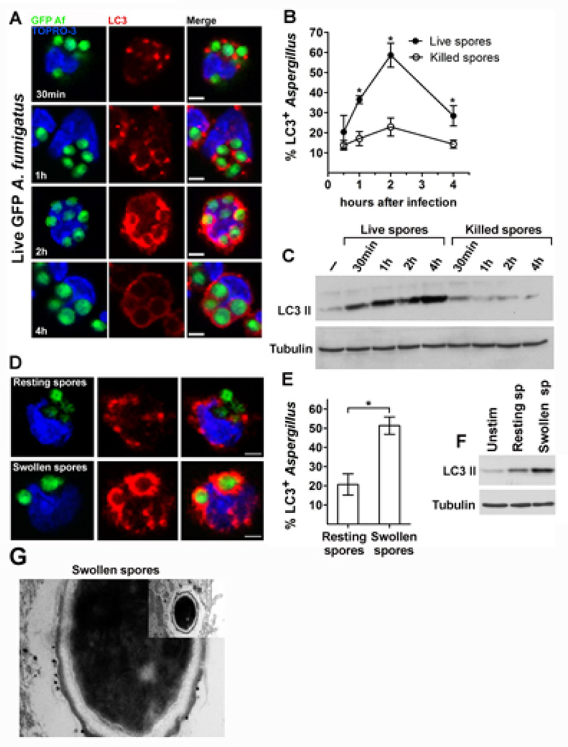
Figure 1. LC3 II is selectively recruited to phagosomes of primary human monocytes during cell wall swelling ofA. fumigatus.
A-B. Primary human monocytes (2 × 10⁵ cells/condition) isolated from healthy individuals were infected with live GFP A. fumigatus (GFP Af; A, B) or PFA-killed GFP A. fumigatus (B) at a MOI 5: 1 for the indicated times. Cells were fixed, permeabilized, stained for LC3 II with the use of an Alexa555 secondary antibody (red) and TOPRO-3 (blue, nuclear staining) and analyzed by immunofluorescence confocal microscopy. The percentages of LC3-associated A. fumigatus-containing phagosomes (LC3+Aspergillus; n > 150 per group) at all time points were quantified by measuring the number of LC3+Aspergillus-containing phagosomes out of the total number of engulfed Aspergillus spores and data are presented as mean + S.E.M. of 3 independent experiments. *, P < 0.0001, paired Student’s t test. Bar, 5 µm. C. Primary human monocytes (2 × 10⁶ cells/condition) were infected with live GFP A. fumigatus or PFA-killed GFP A. fumigatus as in A-B for the indicated times. Cell lysates were prepared and levels of LC3 II protein were determined by immunoblotting. Levels of tubulin in the same lysates were determined by immunoblotting as loading controls. D-E. Primary human monocytes were stimulated for 1h with PFA-killed dormant or PFA-killed swollen spores of GFP A. fumigatus, fixed and stained as in A. The percentages of LC3+A. fumigatus-containing phagosomes (LC3+Aspergillus; n > 150 per group) were quantified and data are presented as mean + S.E.M. of 5 independent experiments. *, P < 0.0001, paired Student’s t test. Bar, 5 µm. F. Primary human monocytes (2 × 10⁶ cells/condition) were left untreated (unstim) or stimulated with either PFA-killed dormant or PFA-killed swollen spores of A. fumigatus. Cell lysates were prepared and LC3 II and tubulin protein levels were determined by immunoblotting. G. Representative immunoelectron micrograph in which LC3 II was labeled in primary human monocytes stimulated for 1h with PFA-killed A. fumigatus swollen spores with 1.4-nm gold particles.

