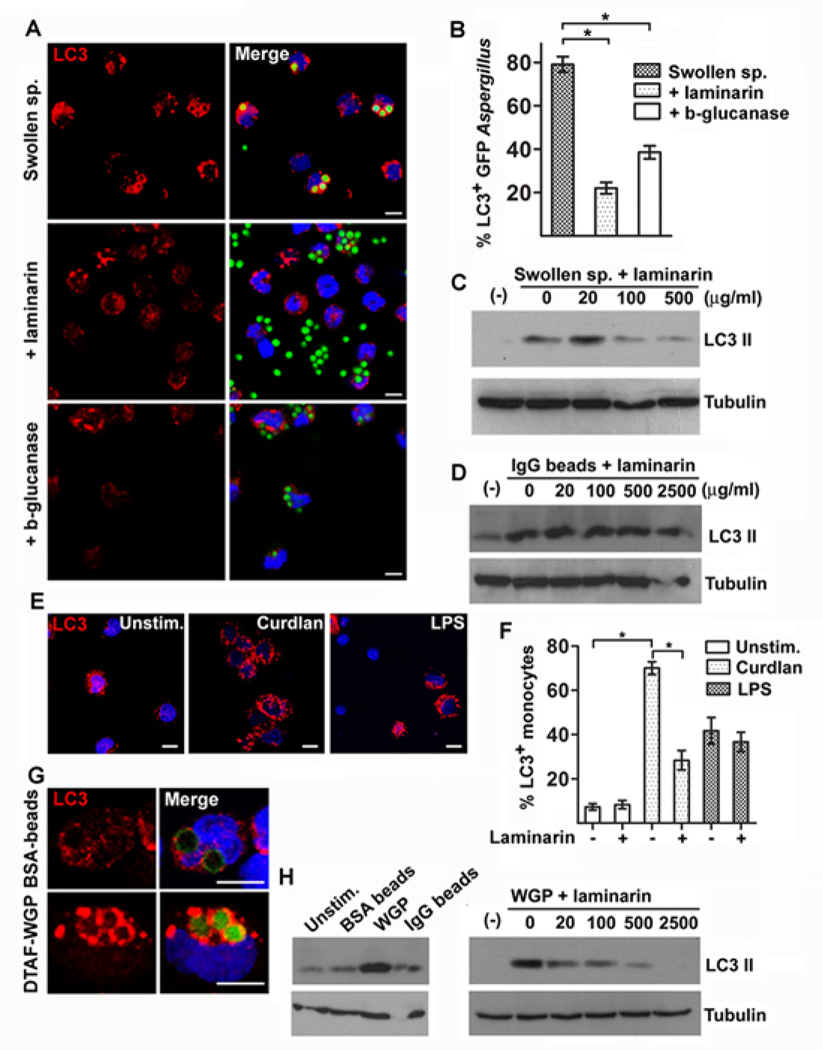
Figure 2. β-glucan surface exposure in swollen spores of A. fumigatus triggers LC3 II recruitment in fungal phagosomes.
A. Primary human monocytes (2 × 10⁵ cells/condition) isolated from healthy individuals were infected with GFP A fumigatus swollen spores with or without laminarin (500 µg/ml) or swollen spores following overnight enzymatic digestion of β-glucan (β-glucanase) at a MOI 5: 1 for 1h. Cells were fixed, permeabilized, stained for LC3 II with the use of an Alexa555 secondary antibody (red) and TOPRO-3 (blue, nuclear staining) and analyzed by immunofluorescence confocal microscopy. Bar, 5 µm. B The percentages of LC3+A. fumigatus-containing phagosomes (LC3+Aspergillus n > 150 per group) were quantified and data are presented as mean + S.E.M. of 3 independent experiments. *, P < 0.0001, paired Student’s t test. C Primary human monocytes (2 × 10⁶ cells/condition) were stimulated with A. fumigatus swollen spores alone or in the presence of increasing concentrations of laminarin, or (D) IgG coated 3mm latex beads alone or in the presence of increasing concentrations of laminarin for 1h. Cell lysates were prepared and levels of LC3 II protein were determined by immunoblotting. Levels of tubulin in the same lysates were determined by immunoblotting as loading controls. E-F. Primary human monocytes (2 × 10⁵ cells/condition) were left untreated or stimulated with purified β-gucan (curdlan, 100 µg/ml) or LPS (100 ng/ml) with or without pretreatment with laminarin (500 µg/ml). The percentages of human monocytes containing autophagosomes as indicated by punctuate LC3 staining (LC3+ monocytes; n > 150 per group) were quantified and data are presented as mean + S.E.M. of 2 independent experiments. *, P < 0.0001, paired Student’s t tes. Bar, 5 µm (G). Primary human monocytes (2 × 10⁵ cells/condition) were stimulated with FITC-labeled BSA beads or DTFA-labeled WGP at a MOI 5: 1 for 1h. Cells were processed as in A and analyzed by immunofluorescence confocal microscopy. Bar, 5 µm. (H). Primary human monocytes (1 × 10⁶ cells/condition) were left untreated or stimulated with BSA coated-beads, IgG coated-beads or WGP with or without pretreatment with increasing concentration of laminarin at a MOI of 10:1 for 1h. Cell lysates were prepared and levels of LC3 II and tubulin were determined by immunoblotting.

