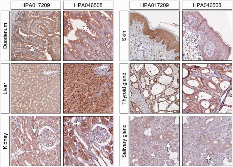
Figure 3.
ERV3 Env protein expression in epithelial cells in the gut (duodenum), liver, skin and thyroid gland. There is good concordance between the two antibodies although the intensity of the staining can vary. The tubular cells of the kidney are moderately stained, but we see very little staining in the glomerular cells. In the salivary glands, the staining is mainly seen in the epithelial cells of the ducts.

