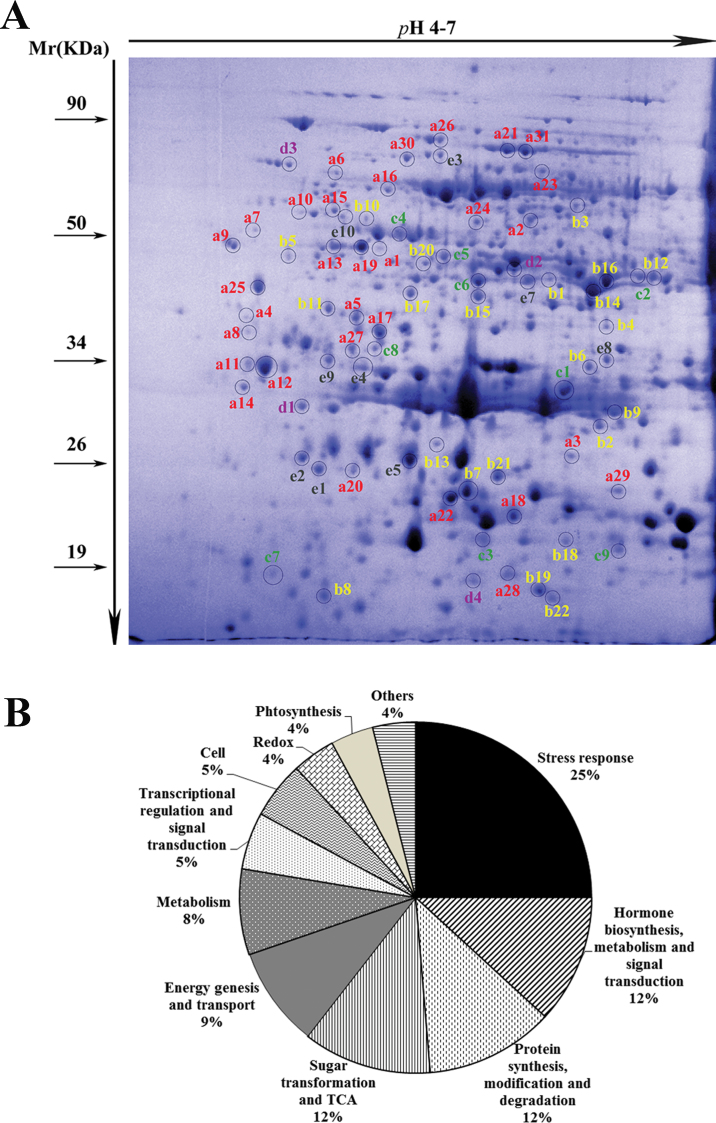
Fig. 6.
Differentially expressed proteins. (A) Image of a representative 2-D polyacrylamide gel. The spots in circles are the identified differentially expressed proteins. The letters a, b, c, d, and e represent five different expression modes: a represents proteins accumulated at one or more time points; b represents proteins that decreased at one or more time points; c represents proteins that accumulated at 12 or 24 HPT and then decreased at 48 or 72 HPT; d represents proteins that decreased at 12 or 24 HPT and then increased at 48 or 72 HPT; and e represents proteins with complex expression modes. (B) Functional categorization and frequencies of all identified proteins in response to 2,4-D treatment. (This figure is available in colour at JXB online.)

