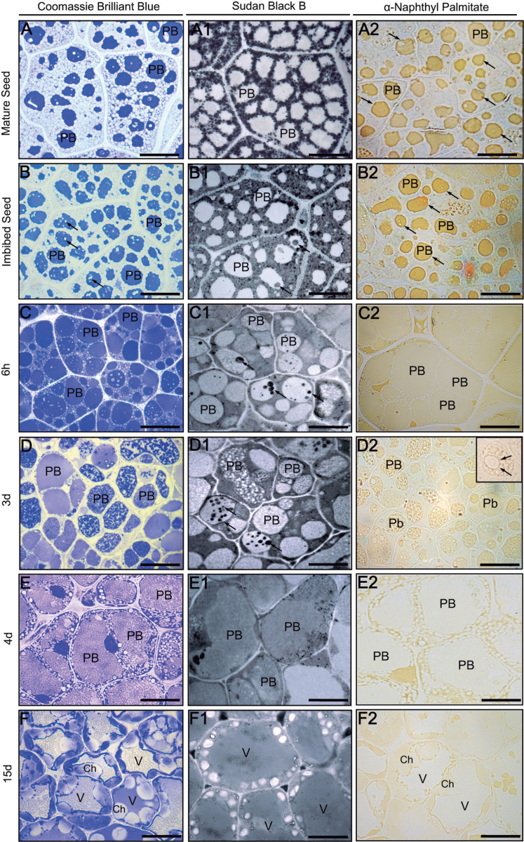
Fig. 3.
(A–F) Coomassie Brilliant blue (CBB) staining of total proteins in sections from olive cotyledons. (A1–F1) Sudan black B (SBB) staining of neutral lipids in sections from olive cotyledons. The arrows denote lipidic masses localized in the protein bodies. (A2–F2) Cellular localization of lipase activity in sections from olive cotyledons. Ch, chloroplast; PB, protein body; V, vacuole. Bars=25 μm.

