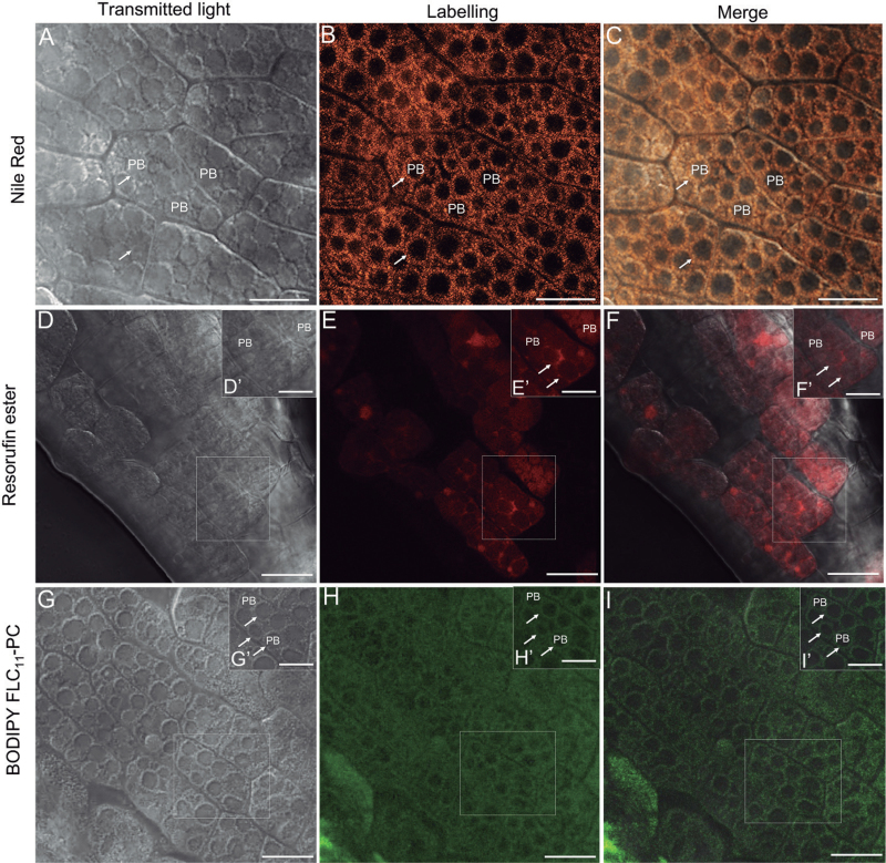
Fig. 7.
Localization of OB lipase (resorufin ester) and phospholipase A (BODIPY® FL C11-PC) activities in living olive cotyledons dissected out from imbibed seed by confocal laser scanning microscopy. (A–C) Optical section of olive cotyledons stained with Nile Red. Numerous OBs are indicated with arrows. (D–F) Olive cotyledons incubated with resorufin ester. Lipase activity is detected in the area of PBs as well as on the OB surface (arrows). (G–I) Olive cotyledons incubated with BODIPY® FL C11-PC. Green fluorescent labelling was detected only on the OB surface (arrows). PB, protein body. Bars=25 μm, inset bars=10 μm.

