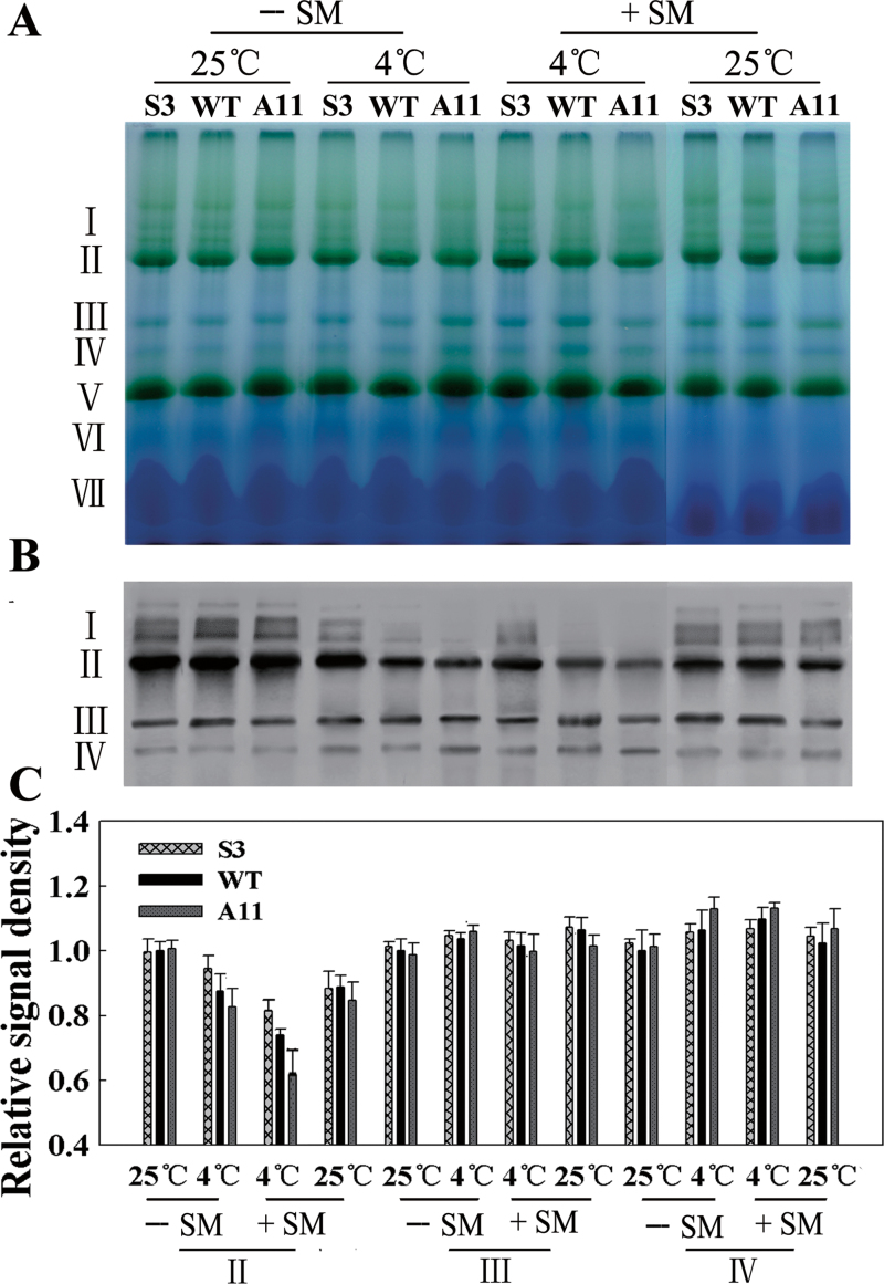
Fig. 10.
Stability of PSII complexes analysed by BN-PAGE and western blotting. (A) BN-PAGE gel. Thylakoid membranes (10 μg of chlorophyll) were solubilized with 1% n-dodecyl β-d-maltoside and separated by BN gel electrophoresis. The positions of protein complexes representing PSII–LHCII supercomplexes (band I), monomeric PSI and dimeric PSII (band II), monomeric PSII (band III), CP43-free PSII (band IV), trimeric LHCII/PSII reaction centre (band V), monomeric LHCII (band VI), and unassembled proteins (band VII). SM was added (+) or not (–) to a final concentration of 3mM. (B) Immunodetection of thylakoid protein complexes separated by BN-PAGE with anti-D1 antibody. The protein complexes were denatured with 15% methanol for 15min before being electroblotted onto PVDF membranes. (C) Quantitative image analysis of the protein content in (B) using a Tanon Digital Gel Imaging Analysis System. The relative protein level of D1 was normalized to that in the WT grown at 25 °C in the absence of SM. Two-month-old grown plants were used.

