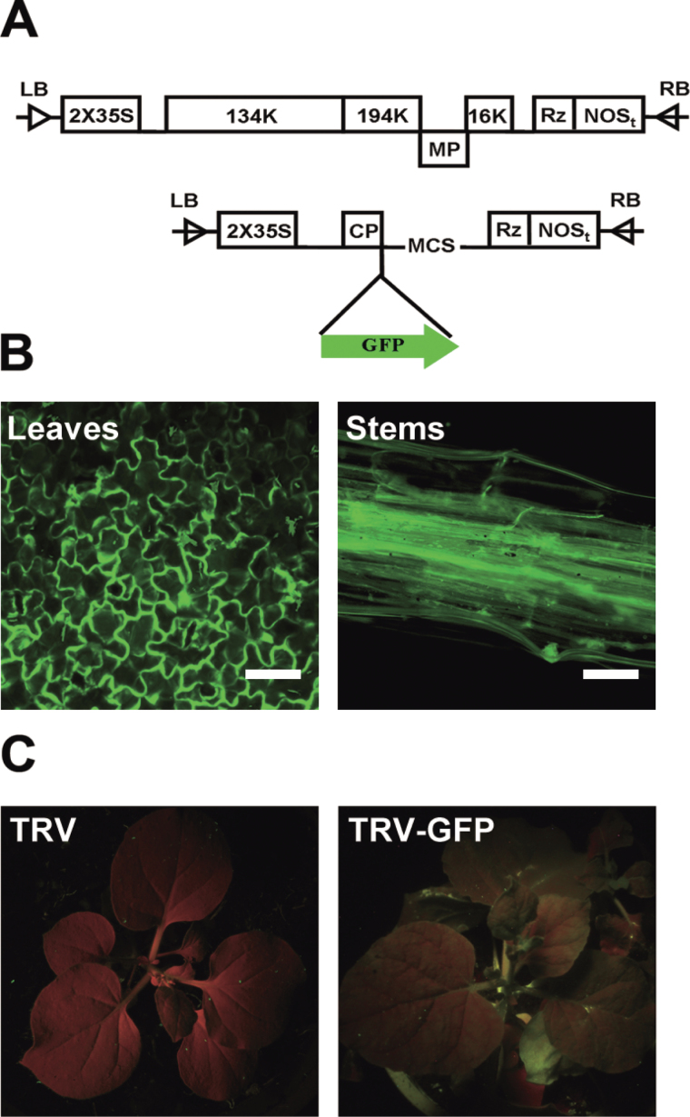
Fig. 1.
Construction and validation of the TRV–GFP vector. (A) Schematic diagram of the TRV–GFP vector. (B) GFP imaging of TRV–GFP-infiltrated N. benthamiana by fluorescence microscopy. (C) Imaging of TRV- (left) and TRV–GFP- (right) infiltrated N. benthamiana by a UV lamp. The TRV 5′- and 3′-untranslated regions (UTRs) are indicated by lines. Open boxes, TRV 2×35S promoter, Rz (self-cleaving ribozyme), nopaline synthase terminator (NOSt), and CP. Green arrow, GFP cistron,. Scale bar=100mm.

