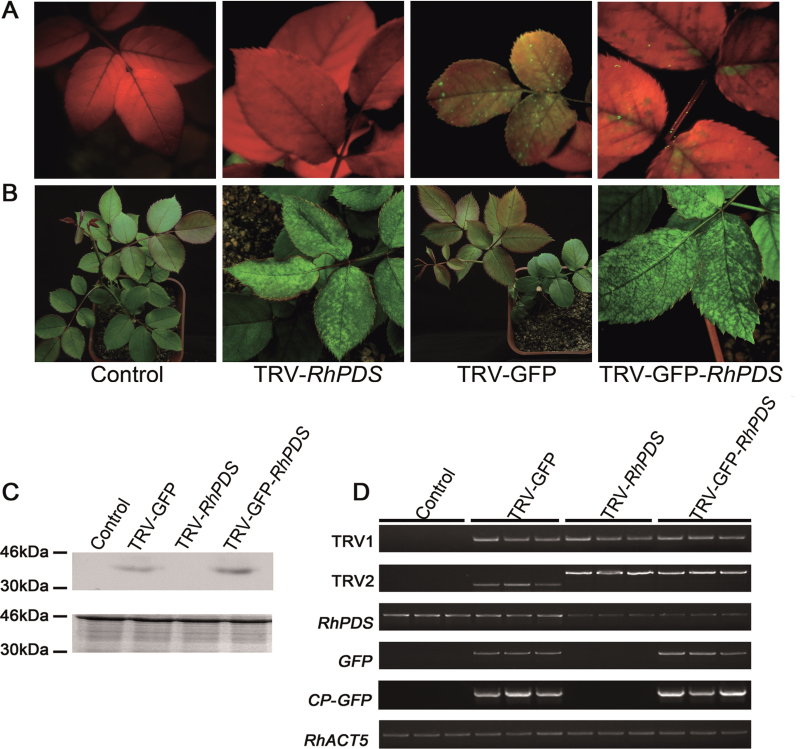
Fig. 6.
Silencing of RhPDS in rose cuttings. (A) TRV- and TRV–GFP-infiltrated rose plants. Rose cuttings were inoculated with a mixture of pTRV2-RhPDS/pTRV2-GFP-RhPDS and pTRV1 (1:1, OD600=1.0) using vacuum infiltration. The plants were photographed under UV illumination and normal light at 30 and 45 dpi, respectively. (B) CP–GFP protein levels in the upper leaves 30 d after inoculation. A 10 μg aliquot of protein was used for western blot in each lane and anti-GFP was used to detect CP–GFP fusion protein. Coomassie blue staining was used for confirmation of equal loading in each lane. (C) Semi-quantitative RT–PCR of TRV1, TRV2, RhPDS, GFP, and CP-GFP in control and inoculated plants. RhACT5 was used as an internal control. Total RNA and protein were extracted from the top young leaves at 45 dpi, which did not exist when the cuttings were infiltrated and grew out at ~15 dpi.

