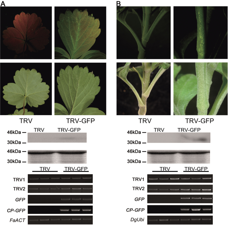
Fig. 9.
Application of TRV–GFP in strawberry (A) and chrysanthemum (B). The plants were inoculated by needleless vacuum infiltration (for strawberry) or syringe injection (for chrysanthemum). The plants were photographed at 21 dpi (for strawberry) and 14 dpi (for chrysanthemum). A 10 μg aliquot of protein was used for western blot in each lane and anti-GFP was used as antibody to detect CP–GFP fusion protein. Coomassie blue staining was used for confirmation of equal loading in each lane. The FaACT and DgUbi genes were used as internal controls for RT–PCR in strawberry and chrysanthemum, respectively.

