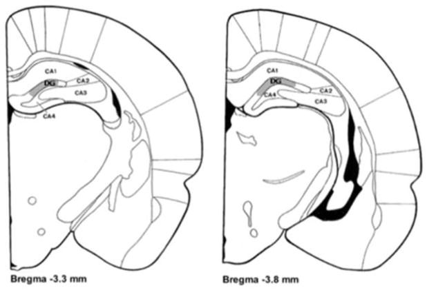
FIGURE 1.
Coronal sections [adapted from Zilles (1985)] showing the region from which dentate granule cells were drawn. Sampling occurred in the upper blade of the Dentate Gyrus (DG), highlighted in gray, in sections ranging from 3.3 mm to 3.8 mm posterior to Bregma.

