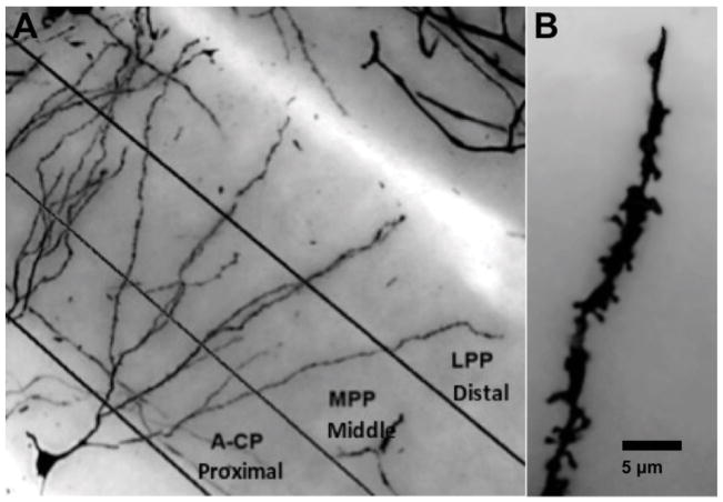
FIGURE 2.
(A) For spine analysis the dentate granule cell dendritic field was divided into 3 layers (proximal, middle, and distal) to correspond to major afferent pathways: A-CP (Associational-Commissural Pathway), MPP (Medial Perforant Path), and LPP (Lateral Perforant Path). (B) Example illustrating the staining quality of granule cell spines (1200X final magnification).

