Figure 5.
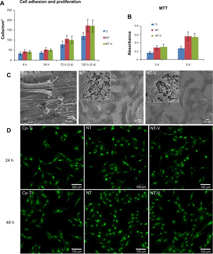
(A–D) Cell behavior on commercially pure titanium (Cp-Ti), TiO2 nanotubes (NTs), and NTs loaded with vancomycin (NT-V). (A and B) Cell adhesion was increased on NTs (both NT and NT-V) compared with the Cp-Ti at 6 and 24 hours (h). At 3 and 5 days (d), the cell numbers had a higher proliferation on the NTs and NT-V compared with the Cp-Ti. (B)The cell proliferation was assessed using MTT-based methods at different time of incubation on the different substrates. (C) Scanning electron microscopy (SEM) micrographs of osteoblasts on Cp-Ti, NT, and NT-V surfaces after 3 days of culture. Higher magnification of the SEM micrographs of osteoblasts on Cp-Ti, NT, and NT-V surfaces showed a much more pronounced protrusion of filopodia, with a significantly longer configuration and a high degree of contact on the NTs (both NT and NT-V) compared to those of the Cp-Ti. The filopodia were also protruding into the nanotube holes on the NTs and NT-V. (D) Fluorescence micrographs of the osteoblast cells after 24 and 48 hours of culture with Cp-Ti, NTs, and NT-V. Living cells (green) and dead cells (red) were stained with acridine orange/ethidium bromide and were visualized using fluorescence microscopy.

