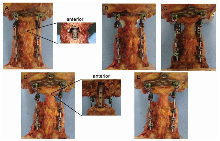
Figure 1.
Photographs of cadaveric occipitocervical constructs tested. (A) C2 corpectomy with cage spanning from anterior arch of C1 to the body of C3, posterior view and anterior view, (B) Complete spondylectomy of C2 with Steinman pins and methylmethacrylate spanning from C1 lateral masses to the body of C3, (C) Complete spondylectomy of C2 with cages spanning from the C1 lateral masses to the C3 facets (D) Complete spondylectomy of C2 and resection of anterior arch of C1 with cage spanning from the clivus to C3 body, (E) Posterior occipitocervical fixation without any other reconstruction after C2 spondylectomy and C1 anterior arch resection.

