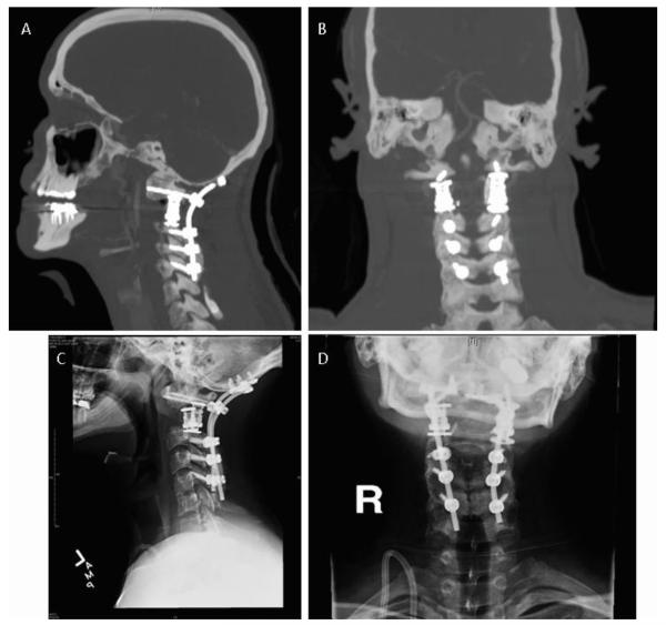
Figure 7.
Postoperative images demonstrating Occiput to C5 fusion with cages spanning the C1 lateral masses to C3 facets. Sagittal (A) and coronal (B) computed tomography reconstructions with patent vertebrobasilar flow on coronal views. Lateral cervical (C) anteroposterior (D) and spine radiographs taken two years post-operatively.

