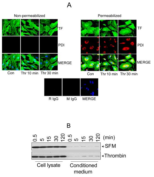
Figure 3. PDI is localized intracellularly in endothelial cells and not secreted in response to thrombin.
(A) Cellular localization of PDI by immunofluorescence confocal microscopy. HUVEC transfected with TF adenovirus were stimulated with thrombin for 10 or 30 min (0.5 U/ml) or left unperturbed. After the treatment, the cells were fixed and permeabilized with 0.25% Triton X-100. Both the intact and permeabilized cells were immunostained with rabbit anti-human TF IgG and monoclonal PDI antibody (RL90), followed by Oregon Green-labeled anti-rabbit IgG and Rhodamine Red-conjugated anti-mouse IgG as secondary reporter antibodies. The bottom panel shows staining with isotytpe controls IgG (B) HUVEC cultured in 24-well plate were treated with 500 μl of control serum-free medium or serum-free medium containing thrombin (0.5 U/ml) for varying time periods. At the end of the treatment, the whole supernatant medium was collected and concentrated by 25 times (to ~20 μl) by ultrafiltration and the cells were lysed in 100 μl of lysis buffer. Equal volumes (10 μl) of cell lysates and the concentrated supernatant media were subjected to SDS-PAGE and immunoblotted with anti-PDI antibodies.

