
Figure 1.
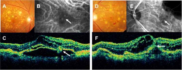
Color fundus photographs (A and D), indocyanine green fundus angiographies (B and E), and optical coherence tomography images (C and F) from the right eye (A–C) and the left eye (D–F) of an 83-year-old woman (patient 1).
Notes: We diagnosed her right eye with stage II retinal angiomatous proliferation and her left eye with stage III retinal angiomatous proliferation. (A and D) Fundus image shows intraretinal hemorrhages with a large number of soft drusen and pigment epithelial detachment. (B) Indocyanine green fundus angiographies shows some hotspots. One of them connects retinal vessels (arrow), corresponding to the intraretinal neovascularization. (C) A vertical optical coherence tomography image shows a pigment epithelial detachment, cystoid macular edema, and retinal angiomatous proliferation lesion (arrow). (E) Indocyanine green fundus angiographies shows choroidal neovascularization (arrow) that connects retinal vessels, corresponding to retinal-choroidal anastomosis. (F) A vertical optical coherence tomography image shows a pigment epithelial detachment, cystoid macular edema, and a retinal pigment epithelium line that has ruptured (arrow).