
Figure 3. Nuclear positioning during cell polarization via TAN lines.
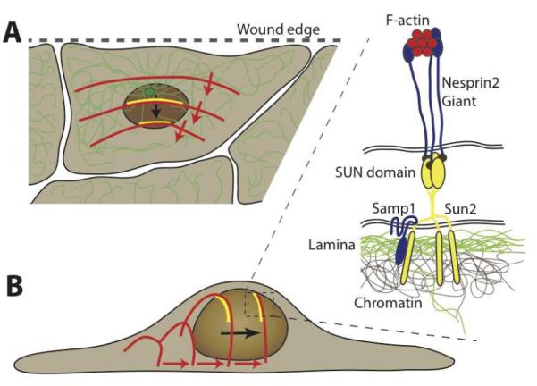
Schematic depiction of retrograde nuclear movement during early polarization in a scratch wound assay. (A) The nucleus moves to the rear end of the cell, resulting in the centrosome (green, with microtubule network) to become located towards the leading edge (i.e., the wound edge) of the cell. Nuclear translocation is mediated by rearward moving dorsal actin cables (red), which form stable connections to complexes of nesprin2, Sun2 and Samp1 (yellow), referred to as TAN lines. (B) Schematic side view of the process by which rearward moving actin cables move the nucleus towards the rear of the cell. The inset shows a close-up of the molecular structure of the TAN lines: F-actin cables interact with the actin-binding domain of nesprin-2 molecules, which bind to Sun2 homotrimers across the perinuclear space. Sun2 also interacts with Samp1 and the underlying nuclear lamina and chromatin.