
Figure 4. Nuclear deformation during cell migration through tight constrictions.
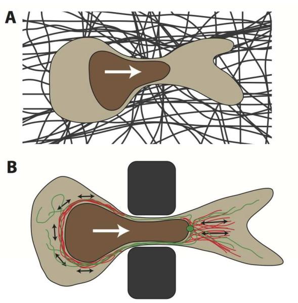
(A) Schematic depiction of a cross-section of a cell migrating through a constriction in the dense extracellular matrix (dark fibers) that is smaller than the nuclear diameter. The white arrow denotes the direction of cell migration. The nucleus is depicted in brown. (B) Sideview of a cell migrating through a polycarbonate filter or microfabricated device used to study nuclear deformation during cell migration through precisely defined pores. Illustrated in red are actinmyosin networks, applying contractile forces (black arrows) to the nucleus, either posterior to the nucleus, resulting in a pushing force, or anterior, pulling on the nucleus. Molecular motors on the microtubule network (green, with centrosome) may apply additional forces to the nucleus, particularly during neuronal migration. White arrow indicates migration direction.