
Figure 8. Flow cytometric analysis of mitochondrial membrane potential in HDF, exposed or not exposed to UVA (365 kJ/m2), determined using the JC-1 assay.
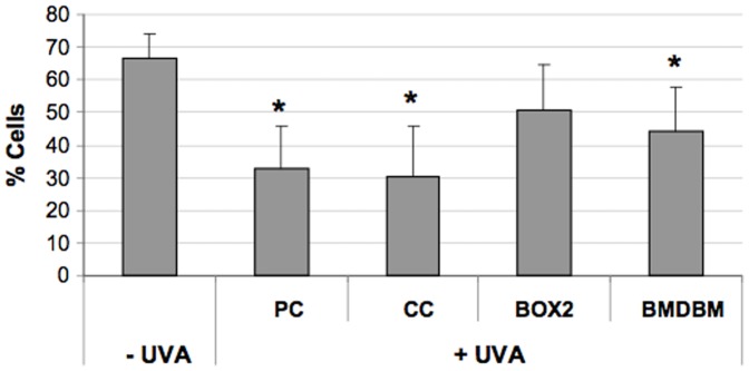
HDF were either screened with formulations (BOX2, BMDBM), control cream (CC) or not screened at all (PC, positive control). Data are reported as percentage of cells presenting high mitochondrial membrane potential in terms of JC-1 red fluorescence. Error bars represent ± S.D. * vs −UVA.