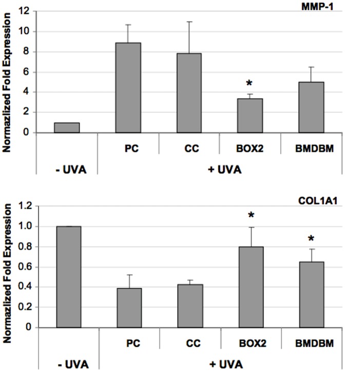
Figure 10. Gene expression analysis of MMP-1 and COL1A1 in HDF, exposed or not exposed to UVA (275 kJ/m2), assessed using qPCR.
HDF were either screened with formulations (BOX2, BMDBM), control cream (CC) or not screened at all (PC, positive control). Data are reported as normalized fold expression using the 2−ΔΔCt method. Error bars represent ± S.D. * vs PC.

