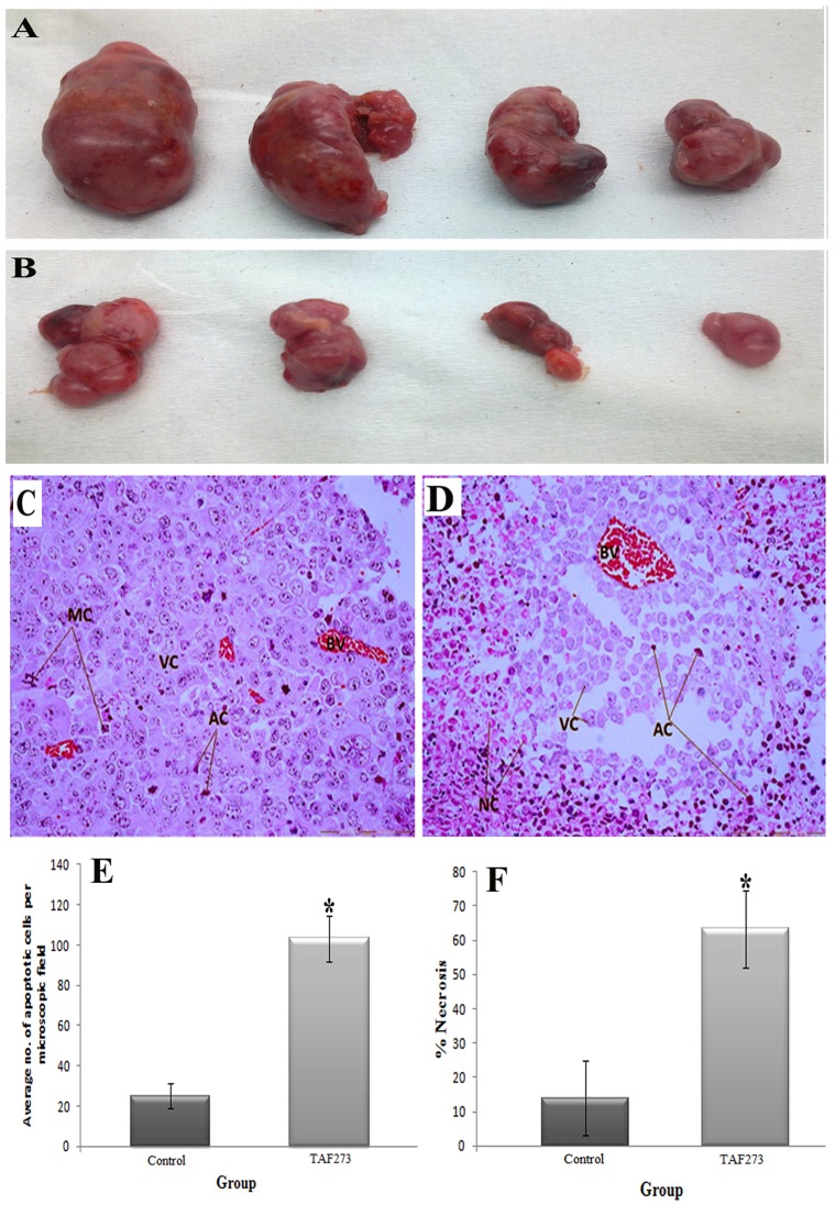
Figure 7. Effect of TAF273 on the size and histological appearance of subcutaneous tumor induced by injecting the K-562 cells in nude mice.
(A) Gross appearance of tumors in the control mice. (B) Gross appearance of tumors in TAF273-treated mice. (C): An H&E-stained tumor section (original magnification of 40×) of the control group is composed of compact sheet of aggressively proliferating viable tumor cells (VC), abundance of blood vessels (BV), and the presence of mitotic figures (MC). (D): The tumor section (original magnification of 40×) of TAF273 (50 mg/kg) IP- treatment revealed notable changes in tumor histology, as significant loss of compact arrangement of viable tumor cells (VC), with less number of blood vessels (BV), abundance of apoptotic cells (AC) surrounded by necrotic regions (NC) and absence of mitotic figures. (E): Graphical comparison of the mean apoptotic cells/microscopic field (control vs TAF273). (F): Graphical comparison of the mean necrotic areas (control vs TAF273) as calculated by using imageJ softwere. Values are presented as mean ± SD, (n = 4).

