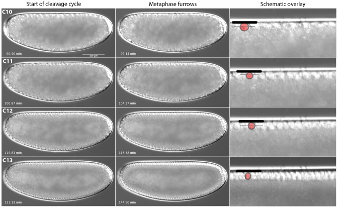
Figure 6. M. abdita early blastoderm cycles (C10–13).
Captured images from live DIC movies. Images show lateral views, anterior is to the left, dorsal is up. Times in min after egg laying (AEL). Schematic overlays show vitelline membrane (thick black line), nuclei (red circle) and metaphase (pseudo-cleavage) furrow front (thin black line).

