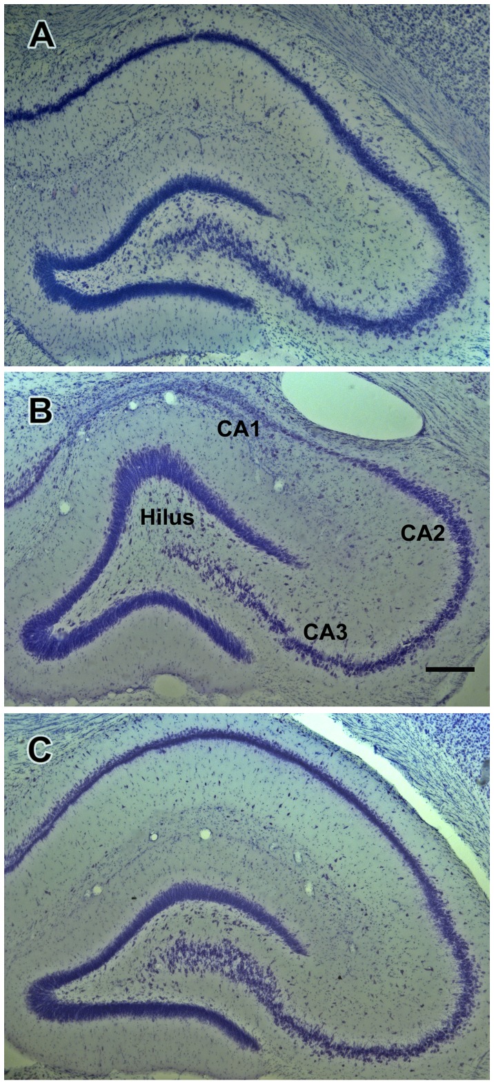
Figure 5. Effects of kainate treatment on hippocampal morphology.
Photomicrographs of representative Nissl-stained coronal sections containing the dentate gyrus, CA3 and CA1 hippocampal fields taken from a control rat (A), from a rat in the behavioral status epilepticus (BSE) group (B) and from a kainate-treated rat that had not experienced behavioral status epilepticus (no-BSE; C). Note that the density of neurons in the dentate hilus is considerably reduced in the rats with (B) and without (C) behavioral SE when compared to the control rat (A). Note also dramatic loss of the CA3 and especially CA1 neurons in the rat from the BSE group; neurons in the CA2 field are relatively preserved (B). Scale bar shown in (B) = 200 µm.

