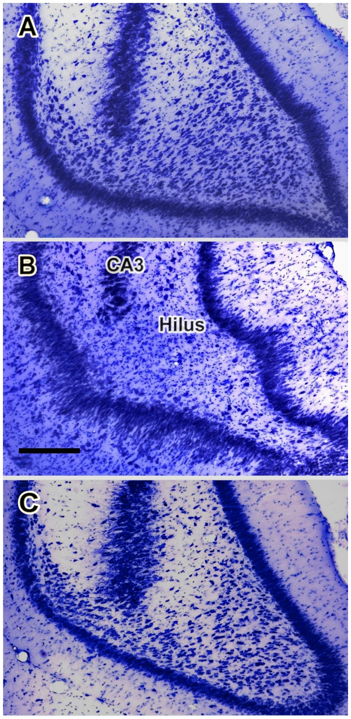
Figure 6. Kainate-induced loss of neurons in the ventral dentate hilus.
Photomicrographs of representative Nissl-stained coronal sections containing the ventral portion of the dentate gyrus taken from a control rat (A), from a rat in the behavioral status epilepticus (BSE) group (B) and from a kainate-treated rat that had not experienced behavioral status epilepticus (no-BSE; C). Note that the density of neurons in the dentate hilus is dramatically reduced in the rat that experienced behavioral SE (B). In striking contrast, most of the neurons of the ventral hilus are preserved in the rat from the no-BSE group (C). Scale bar shown in (B) = 250 µm.

