
Figure 2. Unusual configuration of the HLA-DP2 peptide binding groove.
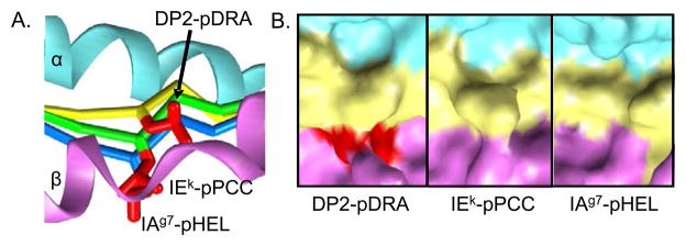
A, A side view of the DP2 β1 helix (magenta) toward the α1 helix (cyan) is shown. The pDRA backbone is shown as a Cα trace (yellow). Two other MHCII/peptide structures, which also have leucines at p4, were overlaid on the DP2-pDRA structure: IEk-pPCC (PDB ID code 1KTD) and IAg7-pHEL (PDB ID code 1F3J). Their peptide Cα traces are shown in green and blue, respectively. For all three peptides, the side chain of the p4Leu is shown as a red wireframe. B, For the same three structures shown in A, the solvent exposed surface of the MHC/peptide complex is shown in the region of p4Leu, α1 - cyan, β1 - magenta and peptide - yellow, expect for the side chain of p4Leu -red.