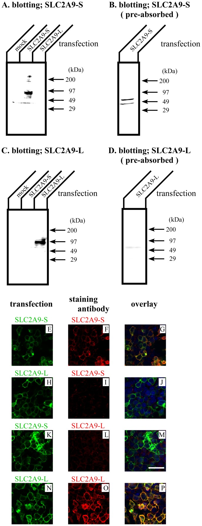
Figure 2. Characterization of isoform specific SLC2A9 antibodies.
30 µg of COS cell lysate transfected with mock or SLC2A9s were separated by SDS-PAGE and Western blotting was performed with anti-SLC2A9-S (A), antigen pre-absorbed anti- SLC2A9-S (B), anti-SLC2A9-L (C), or antigen pre-absorbed anti-SLC2A9-L (D) antibodies. COS cells were transfected with GFP tagged SLC2A9-S (E and K) or SLC2A9-L (H and N) and immunofluorescence was performed with anti-SLC2A9-S (F and I) or anti-SLC2A9-L (L and O) antibodies. Overlay images were shown in G, J, M and P. The scale bar of 40 µm was shown in K.

