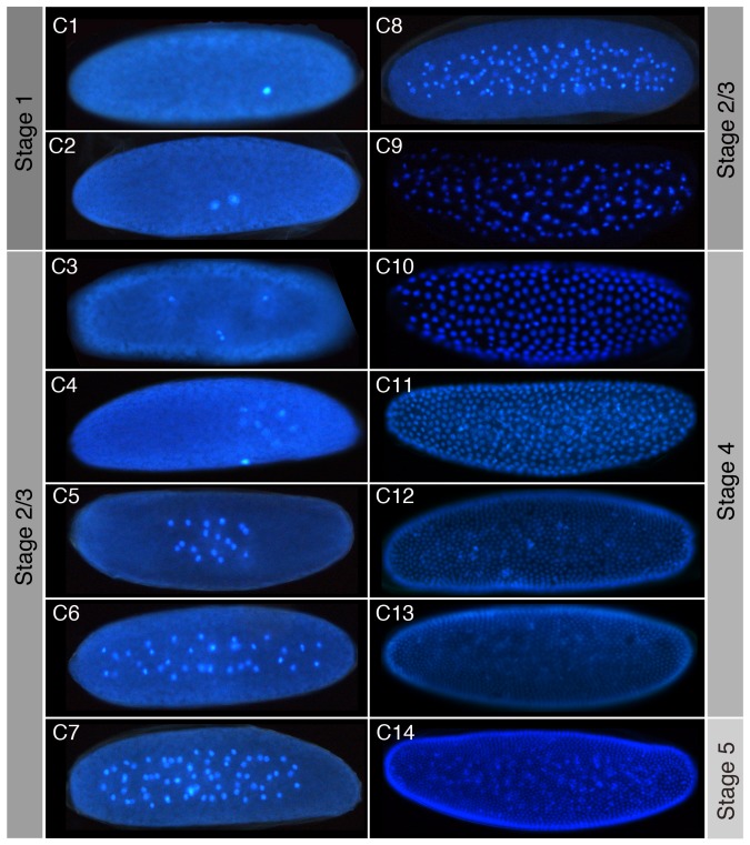
Figure 4. Cleavage cycles of C. albipunctata.
Fluorescence images of embryos with DAPI-counterstained nuclei are shown as lateral views. Anterior is to the left. C1–14 indicates cleavage cycle number. The focus is on the sagittal plane for embryos at cleavage stage (C1–C9), and lateral views are shown at blastoderm stage (C10–14). As in D. melanogaster, nuclei begin to move towards the periphery from C7 onwards. Corresponding embryonic stages (see Figures 2 and 3) are indicated on grey background.

