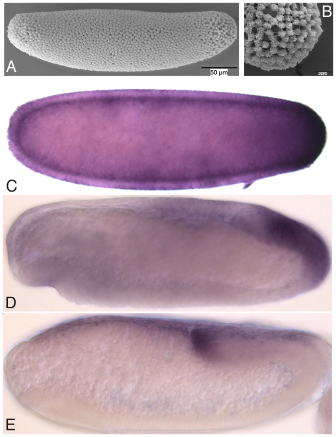
Figure 10. Germ plasm in C. albipunctata.
(A) Scanning electron micrograph of a stage 4 embryo showing the absence of morphologically distinguishable pole cells. (B) Close up of the posterior pole of the embryo shown in (A). (C–E) Vasa antibody stains of embryos at stage 4 (C), 6 (D), and 8 (E). Lateral views, anterior is to the left, dorsal is up. See text for details.

