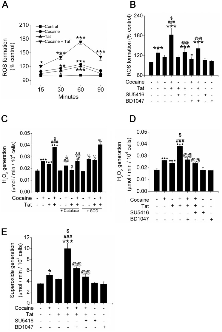
Figure 2. Enhanced oxidative stress on treatment of pulmonary endothelial cells with Tat and cocaine.
(A) Generation of ROS in HPMECs treated with Tat and/or cocaine was quantified by DCF assay at indicated time-points. (B) ROS production in cells treated with Tat and cocaine in the presence or absence of SU5416 (antagonist of VEGFR-2) or BD1047 (antagonist of sigma receptor) for 1 hour as analyzed by DCF assay. (C) Generation of H2O2 was quantified using Amplex red assay kit. HPMECs were treated with Tat and cocaine in the presence or absence of catalase (10U/ml) or SOD (100U/ml) for 1 hour. (D) Reduction of H2O2 formation on pre-treatment of Tat and cocaine exposed HPMECs with SU5416 or BD1047. (E) Changes in Tat and cocaine-mediated superoxide generation on SU5416 or BD1047 pre-treatment. Formation of superoxide (O2 −) was quantified by SOD-inducible cytochrome c reductase assay. The values shown are means (±SD) of at least three independent experiments. *P≤0.05, **P≤0.01, ***P≤0.001 compared to control; #P≤0.05, ##P≤0.01, ###P≤0.001 compared to cocaine treatment; $P≤0.001 compared to Tat treatment; @ P≤0.05, @@ P≤0.001, compared to Tat and cocaine combinational treatment; &P≤0.01, &&P≤0.001 compared to catalase-treated control; %‘P≤0.001, compared to SOD-treated control.

