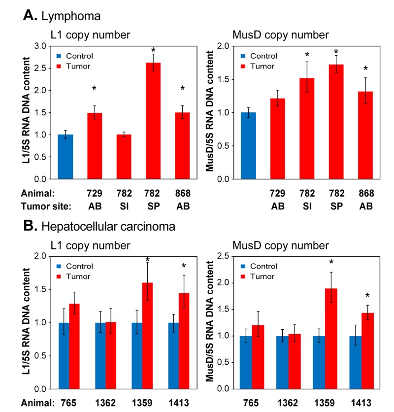
Figure 7. qPCR analysis of DNA to assess RTE genome copy number in spontaneously occurring tumors.
(A) Lymphoma; (B) Hepatocellular carcinoma. Analysis was performed as in Figure 6, except that total DNA was extracted from formalin preserved tissue biopsies (see Methods). Animals are designated by their unique identifier numbers. In (A) the control was normal liver tissue of an animal of comparable age. AB, abdomen; SI, small intestine; SP, spleen. In (B) the controls were normal tissues cut from the vicinity of the tumor. Means and standard deviations are shown. (*) p<0.01.

