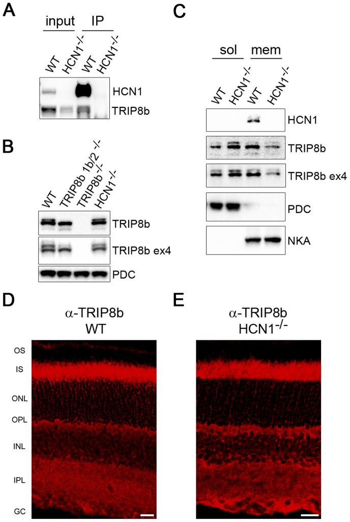
Figure 4. HCN1 is required to fully recruit TRIP8b to the membrane.
A) Anti-HCN1 antibodies were used to co-immunoprecipitate TRIP8b from retinal membranes. Membranes prepared from HCN1−/− retinas were used as the negative control. B) Western blot comparing the amount of TRIP8b present in total retina lysates from wild type, both TRIP8b knockout lines, and HCN1−/− mice. Phosducin (PDC) is the loading control. C) Retina lysates from wild type and HCN1−/− mice separated into cytosolic and membrane fractions probed with anti-TRIP8b and anti-HCN1 antibodies. PDC and sodium/potassium ATPase (NKA) are loading controls for each fraction. Immunostaining of TRIP8b in wild type (D) and HCN1−/− retina (E) is indistinguishable. Abbreviations as in Figure 1; Scale bars are 20 µm.

