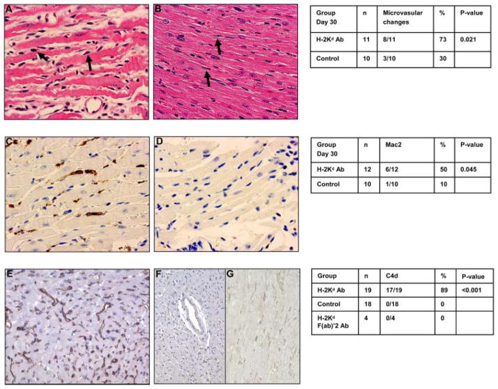
FIGURE 2.
Treatment with anti-donor H-2Kd Ab induces histological features of AMR. BALB/c cardiac allografts were transplanted into B6.RAG1 KO recipients and passively transfused with anti-H-2Kd Ab or mIgG Ab and grafts were examined on days 15 and 30. Microvascular abnormalities were observed by H&E staining in recipients treated with anti-H-2Kd Ab. Arrows indicate prominent nuclei of cells in distended capillaries (A) not seen in isotype control mIgG Ab where arrows indicate flat and thin nuclei in collapsed capillaries (B) on day 30. Intravascular macrophages were assessed by MAC2 immunoperoxidase staining in recipients treated with anti-H-2Kd Ab (C) or isotype control mIgG Ab (D) on day 30. Complement activation was measured by C4d deposition in recipients treated with anti-H-2Kd Ab (E), isotype control mIgG Ab (F) or anti-H-2Kd F(ab′)2 Ab (G). Grafts represent C4d staining on day 30.

