Figure 3.
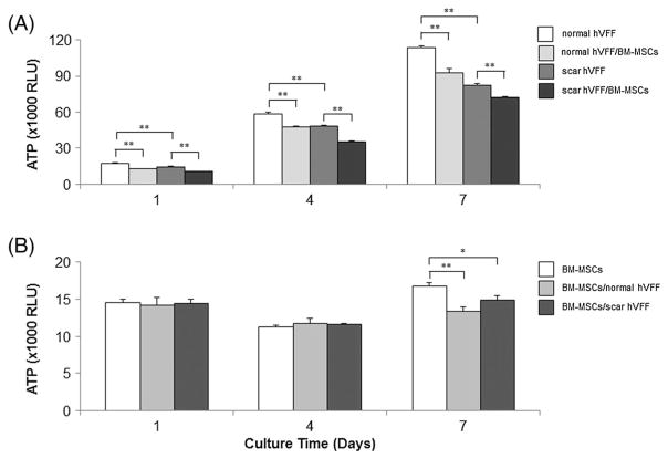
Effect of co-culture on cell proliferation. VFFs were seeded at 10 000 cells/well and grown for 4 h prior to co-culture with BM-MSCs (100 000 cells/well in 3D HA). On days 1, 4 and 7, viable cell numbers of VFFs and BM-MSCs were separately evaluated by ATP amount (RLU) in quadruplicate. Data shown represent mean ± SD of a single representative experiment. (A) ATP levels of VFFs (normal and scarred VFFs) in monoculture and BM-MSCs co-culture conditions; (B) ATP levels of BM-MSCs in monoculture and co-culture conditions. *p < 0.05; **p < 0.01

