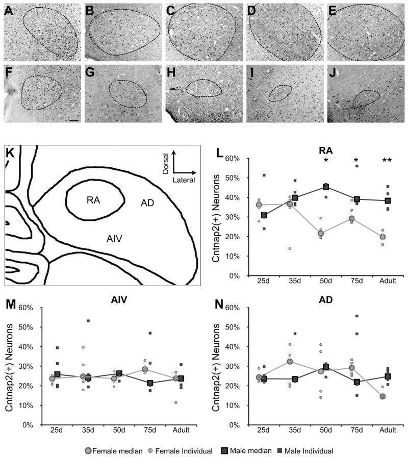
Figure 5. Cntnap2 within RA in both sexes at developmental time points during male song learning.
A-J) Representative images of Cntnap2 immunolabeling of cells in male (A-E) and female (F-J) RA at time points during development encompassing the onset of sensory acquisition, sensorimotor learning, and crystallization of song. Anti-NeuN signals (not shown) were used to trace the border of RA in each image. As previously reported (Konishi and Akutagawa, 1985; Nixdorf-Bergweiler, 1996), the size of RA begins to decrease in females and increase in males starting around 35d and continues through development until maturity. K) A diagram of RA and the two arcopallial regions in which labeled cells were counted: the ventral intermediate arcopallium (AIV) and the dorsal arcopallium (AD). L-N) Graphs representing the percentage of Cntnap2 positive neurons out of the total number of NeuN positive cells found in RA, AIV, and AD, respectively, for 3-6 birds of each sex at each time point. Statistical significance was determined by resampling ANOVA, followed by individual Student’s T-tests *p<0.05, **p<0.01. Scale bar = 100 μm.

